.
Phoenix X-Ray CT
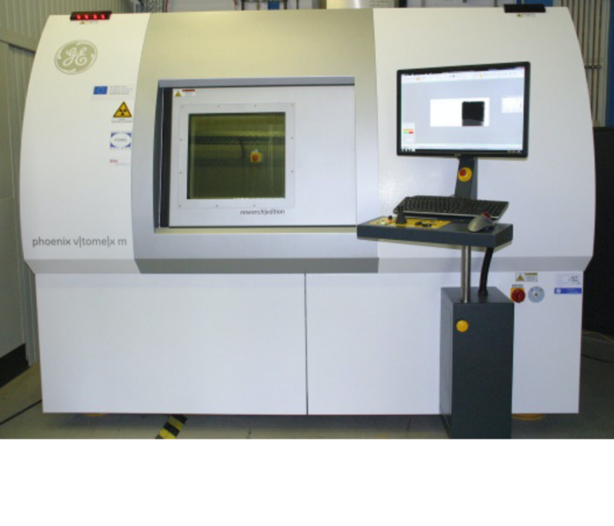
Phoenix-xray v|tome|x m
The scanner
Our Phoenix-xray v|tome|x m is equipped with two X-ray sources: (1) a 180 kV / 15 W nano-focus x-ray tube, and (2) a 240 kV / 320 W micro-focus x-ray tube. For (1), the detail detectability goes down to 1 μm (object size 2 mm), while (2) can penetrate up to 40 mm steel, and has a detail detectability down to 3 μm. Max. object size is (height x diameter) 600 mm x 500 mm, max. object weight is 50 kg.
The method
The method is based on the detection and localisation of the degree of attenuation of the incident X-rays in the sample. The attenuation of X-rays by matter depends on both the chemical elements it consists of as well as the material density. The information about the varied X-ray absorption is encoded as grey values in a stack of black-and-white images. Each image consists of volumetric pixels and contains true volumetric information.
Sample types and preparation
X-ray micro tomography can characterize all materials that are transparent for X-rays, e. g. metals, ceramics, and any kind of sythetic material. The sample surface does not have to be prepared in advance. In our lab we analyze compound materials, opto-electronic components, metals, and more.
mehrSample size and - geometry
In general, samples of any shape can be investigated, however a cylindrical shape is most favorable. Sample size may range from a few millimeters to several centimeters. Most important for a successful scan is a sufficient transmission of the sample for X-rays. It is possible to scan a selected sector of a sample.
X-ray CT: relation of sample size and resolution

What resolution can I expect?
Relevant for the attainable resolution is the position of the sample in the cone beam geometry between the X-ray point-focus source and the detector, which determines the magnification. In general, small samples (1-2 mm) can be characterised with high resolution (<1 - 3 μm per voxel), and larger samples with lower (20-40 μm per voxel).
mehrWhat kind of result do I get?
The result is the reconstructed density distribution within the object in the form of a digital image stack or as image volume, which can be downloaded from the lab server. A storage medium will not be provided. In addition, the transmission images can be supplied. An analysis of the tomography data is possible in the context of a scientific cooperation.
mehrContact
Contact person for technical information
Christian Kapitza
Analytical service
Requests for analyse services are welcome for both academic and non-commercial customers. Please contact
Prof.res Ralf B. Bergmann, Axel Herrmann, or
Christian Kapitza (e-mail: kapitza@bias.de),
Oliver Focke (e-mail: focke@uni-bremen.de).
For usage regulations follow the link:
mehr


